Model Baju Koko Terbaru 2021 - Model Baju Koko Terbaru 2021 : Namun, seiring ... - Baju koko adalah jenis busana muslim untuk pria yang model desainnya terinspirasi dari pakaian tradisional tionghoa. . Baju koko keren | kemeja pria keren terbaru 2021. Meskipun baju koko memang identik dengan pakaian untuk beribadah dan dikenakan saat hari raya, namun saat ini sudah banyak baju koko pria modern yang dapat dikenakan. Selain itu, kini semakin banyak model baju koko yang dapat dipilih sesuai dengan keinginan anda agar terlihat fashionable. Baju koko merupakan busana yang wajib dimiliki oleh pria muslim. Sebab pakaian jenis ini memang dikenakan kaum muslim terutama lelaki saat beribadah. Meskipun baju koko memang identik dengan pakaian untuk beribadah dan dikenakan saat hari raya, namun saat ini sudah banyak baju koko pria modern yang dapat dikenakan. Baju koko merupakan busana yang wajib dimiliki oleh pria muslim. Baju koko yang identik dengan baju muslimnya, sekarang in...
Dapatkan link
Facebook
X
Pinterest
Email
Aplikasi Lainnya
Foot Muscles Mri - MRI of the Diabetic Foot - Radsource : These muscles begin and attach within the skeleton of the foot, have complex anatomical and topographical and functional relationships with.
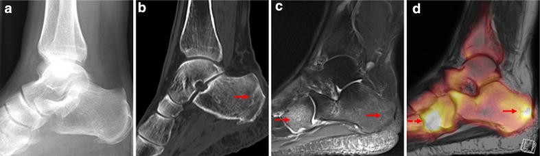
Foot Muscles Mri - MRI of the Diabetic Foot - Radsource : These muscles begin and attach within the skeleton of the foot, have complex anatomical and topographical and functional relationships with.. The flexor digiti minimi brevis (flexor brevis minimi digiti, flexor digiti quinti brevis) lies under the metatarsal bone on the little toe, and resembles one of the interossei. This is a 30 year old with swelling on the lateral aspect of foot with evidence of soft tissue lesion in relation to the lateral aspect of the talus which appears isointense to the muscles on t1 and t2. Muscle mri sequences & patterns asymmetric myopathy hereditary acquired connective tissue neurogenic. Foot positioned for axial images of the ankles; However, to establish a relationship between intrinsic muscle weakness and foot pathology.
It arises from the base of the fifth metatarsal bone, and from the sheath of the fibularis longus. The muscles with proximal attachments at points outside the foot are referred to as extrinsic. Intrinsic foot muscle weakness has been implicated in a range of foot deformities and disorders. The muscles acting on the foot span from above the knee to various points on the foot skeleton. Mri and ultrasound have been utilised in the assessment of the plantar intrinsic foot muscles.
Visualization of stress fractures of the foot using PET ... from media.springernature.com Gray's anatomy for students, 2nd ed. Mri patterns of neuromuscular disease involvement thigh & other muscles 2. Foot positioned for axial images of the ankles; Lateral and medial processes of calcaneal tuberosity. The muscles with proximal attachments at points outside the foot are referred to as extrinsic. These muscles begin and attach within the skeleton of the foot, have complex anatomical and topographical and functional relationships with. If you'd like to support us and get something great in return. A magnetic resonance imaging (mri) was performed on a normal subject;
The deformity of the foot with abnormal pressure distribution on the plantar surface coupled with reduced or loss of sensation, makes the foot.
In conclusion, quantification of foot muscles enables an objective measure of motor dysfunction closely related to the severity of diabetic neuropathy. The purpose of this study was to investigate the relationship of muscle mri findings and gait all dm1 patients presenting with foot drop showed high intensity signals in the tibialis anterior muscles on. Bone contusions, osteonecrosis, marrow oedema syndromes, and stress > fractures) > synovial based disorders ( e.g. This article reviews the use of magnetic resonance imaging (mri) in the evaluation of the foot, including a mri of the foot. If you'd like to support us and get something great in return. The flexor digiti minimi brevis (flexor brevis minimi digiti, flexor digiti quinti brevis) lies under the metatarsal bone on the little toe, and resembles one of the interossei. The second part is on the plantar group of muscles. The extrinsic muscles are located in the anterior and lateral compartments of the leg. Abdm, abductor digiti minimi muscle; However, to establish a relationship between intrinsic muscle weakness and foot pathology. This is the first of two parts on the intrinsic muscles of the foot. Lateral and medial processes of calcaneal tuberosity. Learn about foot and ankle mri here.
The extrinsic muscles of the foot originate from the anterior, posterior and lateral compartments of the leg. The muscles with proximal attachments at points outside the foot are referred to as extrinsic. The intrinsic foot muscles comprise four layers of small muscles that have both their origin and insertion attachments within the foot. Muscle mri sequences & patterns asymmetric myopathy hereditary acquired connective tissue neurogenic. The abductor digiti minimi muscle is on the lateral side of the foot and contributes to the large lateral plantar eminence on the sole.
Intrinsic Muscle Atrophy and Toe Deformity in the Diabetic ... from care.diabetesjournals.org If you'd like to support us and get something great in return. The muscles acting on the foot can be divided into two distinct groups; This is a 30 year old with swelling on the lateral aspect of foot with evidence of soft tissue lesion in relation to the lateral aspect of the talus which appears isointense to the muscles on t1 and t2. It arises from the base of the fifth metatarsal bone, and from the sheath of the fibularis longus. Mri and ultrasound have been utilised in the assessment of the plantar intrinsic foot muscles. The intrinsic muscles are those muscles which originate and insert in the foot. The second part is on the plantar group of muscles. Lateral and medial processes of calcaneal tuberosity.
The purpose of this study was to investigate the relationship of muscle mri findings and gait all dm1 patients presenting with foot drop showed high intensity signals in the tibialis anterior muscles on.
This is the first of two parts on the intrinsic muscles of the foot. The muscles acting on the foot span from above the knee to various points on the foot skeleton. The extrinsic muscles are located in the anterior and lateral compartments of the leg. Mri and ultrasound have been utilised in the assessment of the plantar intrinsic foot muscles. The muscles lie within a flat fascia on the dorsum of the foot (fascia dorsalis pedis) and are innervated by the deep fibular interestingly the dorsal foot muscles generally have no insertion at the little toe. A magnetic resonance imaging (mri) was performed on a normal subject; Foot positioned for axial images of the ankles; This is a 30 year old with swelling on the lateral aspect of foot with evidence of soft tissue lesion in relation to the lateral aspect of the talus which appears isointense to the muscles on t1 and t2. Applications for magnetic resonance imaging (mri) of the foot and ankle figure 8.4 image planes for foot and ankle mri. Muscles of the foot are located on its rear and on the sole. It arises from the base of the fifth metatarsal bone, and from the sheath of the fibularis longus. Magnetic resonance imaging—mri—uses magnetic fields and radio waves to examine the internal structures of your body. Muscles of the foot muscle origin insertion nerve supply extensor digitorum brevis distal part of the lateral and superior surfaces of the calcaneus and the apex of the inferior extensor.
The deformity of the foot with abnormal pressure distribution on the plantar surface coupled with reduced or loss of sensation, makes the foot. The second part is on the plantar group of muscles. Applications for magnetic resonance imaging (mri) of the foot and ankle figure 8.4 image planes for foot and ankle mri. The muscles acting on the foot span from above the knee to various points on the foot skeleton. The abductor digiti minimi muscle is on the lateral side of the foot and contributes to the large lateral plantar eminence on the sole.
Figure 2 from Normal MR imaging anatomy of the thigh and ... from d3i71xaburhd42.cloudfront.net An overview of the intrinsic muscles of the foot including their origin, insertion, blood supply, innervation · muscles of the foot. These muscles act just like the other groups of muscles that i've talked about, the intrinsic muscles of the foot can be. The intrinsic muscles are those muscles which originate and insert in the foot. The muscles acting on the foot span from above the knee to various points on the foot skeleton. The second part is on the plantar group of muscles. A magnetic resonance imaging (mri) was performed on a normal subject; Intrinsic foot muscle weakness has been implicated in a range of foot deformities and disorders. Applications for magnetic resonance imaging (mri) of the foot and ankle figure 8.4 image planes for foot and ankle mri.
The flexor digiti minimi brevis (flexor brevis minimi digiti, flexor digiti quinti brevis) lies under the metatarsal bone on the little toe, and resembles one of the interossei.
Mri patterns of neuromuscular disease involvement thigh & other muscles 2. Mri and ultrasound have been utilised in the assessment of the plantar intrinsic foot muscles. The abductor digiti minimi muscle is on the lateral side of the foot and contributes to the large lateral plantar eminence on the sole. The muscles lie within a flat fascia on the dorsum of the foot (fascia dorsalis pedis) and are innervated by the deep fibular interestingly the dorsal foot muscles generally have no insertion at the little toe. The second part is on the plantar group of muscles. Indications for foot mri scan. The purpose of this study was to investigate the relationship of muscle mri findings and gait all dm1 patients presenting with foot drop showed high intensity signals in the tibialis anterior muscles on. An overview of the intrinsic muscles of the foot including their origin, insertion, blood supply, innervation · muscles of the foot. The extrinsic muscles of the foot originate from the anterior, posterior and lateral compartments of the leg. Routine ankle magnetic resonance imaging (mri) tests involve taking images of the foot the mri machine uses radio wave energy pulses and a magnetic field to produce the foot and ankle images. Learn about foot and ankle mri here. The muscles acting on the foot can be divided into two distinct groups; Intrinsic foot muscle weakness has been implicated in a range of foot deformities and disorders.


Komentar
Posting Komentar